Under the Microscope

ctwo

Active member
May 16, 2019
247
145
Parrots
Mango the Indian Ringneck and Peach the Cockatiel; Kiwi found a new home
Relevant to birds, does anyone know what typical parasite and bacterial infections one can observe under an amateur microscope (1000x), or what can I look for related to stool samples and swabs, and maybe a blood sample if I get something from a discarded feather?

My vet said that the cockatiel "might" have some kind of parasite but it was not conclusive, so something to watch...

This seems like a good start, but looking for moer, maybe even just search terms:

https://www.finchaviary.com/Maintenance/FecalSmear.htm
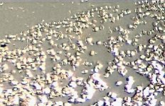
 

charmedbyekkie

New member
May 24, 2018
1,148
82
US/SG
Parrots
Cairo the Ekkie!
Oh, goodness! Do you have an OpenFlexure Microscope too? We've been trying to figure out how to understand our little guy's poop as well :p Apparently the prep of the slide and sample is key to seeing moving things :eek:

My partner and I are by no means biologists, and our friends whom we got advice from are not professional biologists either. But the maker/hacker way we were told was to get the sample, add a small drop of olive oil (or water) (depending on oil or water emulsion), put on the cover slip, seal it up with nail polish so it doesn't dry out, and let it grow first for a day or so. Then check it under the microscope.

Again, not scientific. But if you want scientific, go to your avian vet.

We're just curious about Cairo's poop and seeing the difference between when he eats regular chop and when he eats durian. Yes, it would be helpful to see early on if his poop looks ok and, if it doesn't, then take him to the vet to get tested and diagnosed there. After all, my vet does her faecalysis within 20 minutes :p not the few days that we would do for the homemade microscope.
 
OP
ctwo

ctwo

Active member
May 16, 2019
247
145
Parrots
Mango the Indian Ringneck and Peach the Cockatiel; Kiwi found a new home
  • Thread Starter
  • Thread starter
  • #6
Thanks all for the links and the photos will be useful. I was just looking at a budgie fecal smear and after not seeing anything of interest, I started thinking of the cycle of life and thought I'd have to culture a sample. I wonder how the vets get it done in such short time...?

Well, I do not have an OpenFlexure, but I have had an American Optics inverted microscope for some time just collecting dust, and it only has a max 10x optic with 10x eyepieces. One of the mysteries was getting a size reference of these microbes. I've come across a lot of images of various sorts and many have a scale, but what would those look like in my scope? To answer that, here is a hooman blood sample where one of these cells should be around 7μm. There are only a few cells around the edge that are distinguishable because of the lack of proper preparation. It's literally just a crusted drop on a piece of plastic container, and the photo is much worse than what the eye sees due to poor form (afocal handheld cell phone) and overexposure.

The other day I ordered a better American Optics microscope with more objectives and proper lighting. These seem to be good microscopes for the cost, but nothing to compare with today's lab grade standards.
 

Attachments

  • blood-20191028_205123.jpg
    blood-20191028_205123.jpg
    38.1 KB · Views: 68
Last edited:

Most Reactions

Latest posts

Top